Tag: dna
Stress Reaction May Be In Your Dad’s DNA, Study Finds

Stress Reaction May Be In Your Dad’s DNA, Study Finds

By Geoffrey Mohan, Los Angeles Times (TNS)

Stress in this generation could mean resilience in the next, a new study suggests.
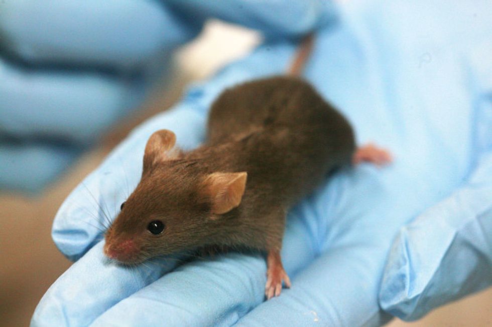
Male mice subjected to unpredictable stressors produced offspring that showed more flexible coping strategies when under stress, according to a study published online Tuesday in the journal Nature Communications.

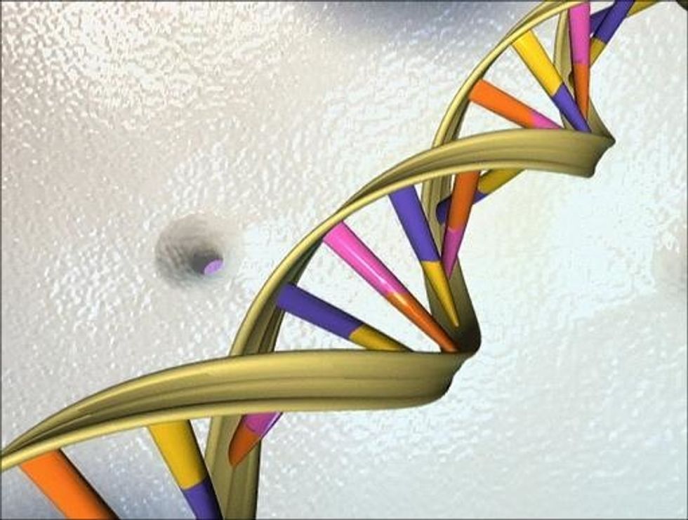
The secret might be hidden in a small change in how certain genes are regulated in the sperm of the father and in the brains of offspring, the study found.

Several studies have shown that stress in early life not only can affect the individual’s behavior and cognitive functions, but can affect the next generation. So researchers have been eager to find any trace of changes in DNA coding that might underlie their observations.

Before you pen a “thanks for the resilience” Father’s Day card, consider: The study involved mice, not humans. More important, even the seemingly more resilient mice had lots of negative behaviors — depression and anti-social tendencies among them.

“If we look at the whole behavior of these animals, the benefit is really a very small proportion of the effects,” said study co-author Isabelle Mansuy, a neuroscientist at the University of Zurich’s Brain Research Institute. “Most other effects are fairly negative, because the animals are depressed, are anti-social, and have cognitive impairment.”

Researchers tried to mimic the effects of erratic parenting and a stressful home environment. So they separated male mouse pups from their mothers for several hours a day over the first two weeks of life, during which time they were occasionally restrained or forced to swim for five minutes — all at unpredictable intervals.

The mice then matured in social groups of four or five unrelated mice of the same sex that had equally unpredictable childhoods. Then they were matched to females, producing pups of their own. Once the pups grew up, they were subjected to various mazes that test the ability to show goal-oriented and flexible behavior under stress.

Compared with a control group, the offspring of stressed dads showed less hesitation in exploring an arm of a maze. And when offered the choice of getting a drink of water immediately or waiting for sugared water, the offspring of stressed males tended to wait for the greater reward. They also were better at figuring out changed rules — rewards that were moved from one spot to another, or cues that were changed.

Numerous studies of the effects of stress implicate a loop in the brain’s limbic system, which mediates emotion and causes the release of the stress hormone cortisol. That chemical can amp up a feedback loop to the brain.

Much of this stress-related reaction in the brain is mediated, in part, by a mineralocorticoid receptor, or MR, in brain cells.

The study found small changes in regulatory DNA sequences near an MR gene in sperm cells of the stressed mice. Such changes in gene regulation in response to the environment are known as epigenetic processes. The study found epigenetic markers associated with a half-dozen genes in the brain cells in the hippocampus of the offspring of stressed male mice.

Together, these changes offer a hint at a possible path for passing the effects of stress from one generation to the next.

Soldiers may offer a prime example, Mansuy said. “Many soldiers are people from lower socioeconomic environments and many of them have been exposed to violence, to broken families and to bad conditions when they were young,” she said. “And many of these people are stress-resilient, and they also have some adaptive advantages when they are placed in a situation of danger or challenge. They have developed coping strategies perhaps that other people have not.”

Still, she noted, these enhanced resiliency behaviors were “the only benefit” observed among the mice.

Researchers have been trying to untangle the effects of genetics and family background in post-traumatic stress disorder among soldiers returning from war.

Photo via WikiCommons

‘Physician-Scientists’ Transform Health Care For Amish, Mennonite

‘Physician-Scientists’ Transform Health Care For Amish, Mennonite

By Amy Worden, The Philadelphia Inquirer

STRASBURG, Pa. — Weeks after his birth in 2001, Benjamin Glick was stricken with a mysterious illness.
He would vomit and pass out. He wouldn’t eat and lost weight. Over five agonizing months, his parents took him to 12 doctors at six hospitals in the Philadelphia area.
“He was fading out, we were going to lose him,” said his father, Amos Glick, who is Old Order Amish and runs a foundry in Chester County.
It took a clinic in a Lancaster County cornfield to save the boy.
Doctors at Children’s Hospital of Philadelphia sent the family to the Clinic for Special Children in Strasburg. For years, the site had been refining an unusual specialty: treating Amish and Mennonite children with rare genetic disorders.
Clinic doctors had seen similar symptoms in other children from the insular population they serve. They discovered Benjamin had a debilitating milk-protein allergy and fine-tuned his formula. Benjamin stabilized in a month.
There was no genetic diagnosis, but two of his siblings born later suffered similar symptoms. Together, their medical histories will provide a foundation for what may lead to a genetic link.
A big hospital likely would have treated each child individually, never making the connection to help those children and others, said Kevin Strauss, a pediatrician and the clinic’s medical director. The case was also meaningful for another reason.
“The modern American medical system,” he said, “didn’t have a place for them to go.”
In the 25 years since its founding, the clinic has transformed pediatric health care for an underserved population, turned diseases from death sentences into treatable conditions, and broken ground in genetics that could one day lead to cures for diseases that afflict the wider population.
The barn-like building, raised in a day by Amish and Mennonite craftsmen, has parking spots for horses and buggies out front, and dairy cows graze out back. Inside, 63-year-old pediatrician D. Holmes Morton and his team practice cutting-edge medicine.
Morton was a Children’s Hospital fellow in 1988 when he encountered a 6-year-old Amish boy with an undiagnosed disease that left him brain damaged and unable to use his limbs.
Doctors thought it was cerebral palsy. Morton, a specialist in biochemical genetics, identified Danny Lapp’s disease as glutaric aciduria type 1 (GA1), a metabolic disease that attacks the brain with sudden and catastrophic results.
At the time, there were only eight known cases.
The Lapp case changed Morton’s life and led to discoveries that have since saved many of the 2,500 patients he has seen. GA1 is one of more than 150 diseases or genetic mutations the clinic has identified that affect the Amish and Mennonites of Lancaster County — but that are not exclusive to them.
The county’s 60,000 Plain People, as they are called, descended from fewer than 100 settlers who came to Lancaster from Europe in the early 1700s. Centuries of intermarriage have increased the risk for developing many conditions.
For instance, Amish babies are 100 times more likely to have GA1 than other infants. At the same time, Morton said, diseases that strike the general population, such as cystic fibrosis and sickle cell disease, are nonexistent among the Plain People.
Before 1990, 90 percent of children suffering from GA1 had irreversible brain damage that left most of them severely disabled. Now, if caught early, those with the disease suffer no brain injury and, with vitamins and dietary restrictions, those the clinic treats have been able to live normal lives.
Vanessa Guimaraes’ 3-year-old daughter was diagnosed with GA1 during a newborn screening. Guimaraes, a native of Brazil living at the time in Hawaii, decided to move to Lancaster to be close to treatment.
“I thank God every day for the clinic,” she said.
Jan Bergen, chief operating officer at Lancaster General Hospital, said she was “awestruck” by the small clinic’s big results.
“They are unique in the world,” said Bergen, who works with the clinic on research.
Born in Fayetteville, W.Va., Morton was a high school dropout before, as he puts it, talking his way into Trinity College. Later, he was accepted at Harvard Medical School.
After Children’s Hospital, Morton decided he wanted to bring medicine to the people but found making inroads with the Amish and Mennonite difficult. A handful, mostly skeptics, came to his first meeting in 1988.
Last month, several thousand Plain People were on hand for a quilt auction to benefit the clinic. Morton rose to thank them for their help, gazing out over a sea of straw hats and prayer caps.
“I think of the clinic as a health care model,” he said in his office later.
In addition to seeing patients every day, clinic researchers publish three to five peer-reviewed papers a year and participate in 25 research projects with hospitals worldwide.
Strauss, the Harvard-trained medical director, said if he had taken a traditional path, he’d be spending his career in a large hospital lab “with mice and fruit flies.” He describes himself today as a physician-scientist, with patient care guiding him.
“We do research on whatever problem comes through the door,” he said.
(EDITORS: STORY CAN END HERE)
Forty percent of the patients diagnosed have treatable — if not curable — conditions. Another 40 percent are partially treatable. The remaining 20 percent have terminal conditions.
Some leave with no diagnosis. That’s where Erik Puffenberger comes in.
On a recent afternoon, Puffenberger, a geneticist and the clinic’s lab director, sat hunched over vials of DNA in a basement lab.
When Morton founded the clinic, he said, he thought he might find 10 to 20 new diseases in his career. Puffenberger identifies five to 10 gene mutations a year.
A freezer contains 8,000 DNA samples from patients that Puffenberger says will provide the foundation for understanding as-yet-unidentified diseases.
He’s working on developing a quicker, cheaper test for Down syndrome among newborns. “Our patients have no insurance and they may be of limited means,” Puffenberger said.
With a staff of 14 and an annual budget of $2.6 million — one-third funded by quilt auctions, the rest by fees and donations — the clinic has been able to offer its largely uninsured patients services at a fraction of a hospital’s cost.
Morton, recipient of the Albert Schweitzer Prize for Humanitarianism and a MacArthur Foundation “genius grant,” is looking to the future. The clinic recently started a fellowship to cultivate the next generation of physician-scientists, and Morton is involved in building a clinic to serve the Amish population near State College.
The treatment Glick’s son Benjamin got 13 years ago made the difference for two of his other six children. Daughters Naomi and Katie, now 7 and 3, were diagnosed quickly and spared any suffering.
“What’s really inspiring to me is that these doctors and scientists could be working in fancy hospitals making six-figure incomes, but they have — as Dr. Puffenberger put it — chosen the road less traveled,” Glick said. “They are making a serious difference in families like ours in our local community and in Brazil and India and Canada from this little place in a cornfield.”

Photo via Wikicommons

Interested in more national news? Sign up for our daily email newsletter!

Forensic Sleuths Sketch Richard III’s Brutal End

Forensic Sleuths Sketch Richard III’s Brutal End

Paris (AFP) — King Richard III likely perished at the hands of assailants who hacked away pieces of his scalp and rammed spikes or swords into his brain as the helmet-less monarch knelt in the mud.

So suggests a report, published Wednesday, that in dry forensic prose exposes the horrific demise of one of English history’s most controversial monarchs.

It backs anecdotal evidence, made famous by Shakespeare, that Richard was unhorsed before he met his doom.

Bringing together 21st-century science and sketchy knowledge of 15th-century history, the analysis provides a chilling tableau of the brutality of warfare in late medieval England.

Richard was killed in the Battle of Bosworth Field in Leicestershire, central England, on August 22, 1485.

The monarch’s death was the culmination of a three-decade war for the throne, bringing the curtain down on the three-century dynasty of his Plantagenet clan, and ushering in the Tudors.

“The most likely injuries to have caused the king’s death are the two to the inferior aspect (lower part) of the skull — a large sharp-force trauma possibly from a sword or staff weapon, such as a halberd or bill, and a penetrating injury from the tip of an edged weapon,” said Guy Rutty, a pathologist at the University of Leicester.

A halberd was a medieval battle ax with spiked point, and a bill was a hooked-tip blade on the end of a pole.

– No horse –

“Richard’s head injuries are consistent with some near-contemporary accounts of the battle, which suggest that Richard abandoned his horse after it became stuck in a mire and was killed while fighting his enemies,” said Rutty.

The study, published in The Lancet medical journal, used X-ray computed tomography (CT) for a microscopic analysis of a skeleton found in 2012 under a car park at a former church.

After being lost for five centuries, researchers identified the remains as Richard’s, backed by DNA analysis and radiocarbon-dating.

The new paper documents nine injuries to the head at or shortly before death, and two to the torso that were likely inflicted post-mortem.

The two blows that probably killed the king likely came from a sword or spike driven into the brain at the base of the skull.

They are consistent with the victim having been “in a prone position or on its knees with the head pointing downwards,” the study’s authors wrote.

Non-fatal injuries included three cuts to the top of the skull that would have sliced off much of the scalp. A knife or dagger was stuck right through his face, from right cheek to left.

“Richard’s injuries represent a sustained attack or an attack by several assailants,” said Sarah Hainsworth, a professor of materials engineering at the university.

“The wounds to the skull indicate that he was not wearing a helmet, and the absence of defensive wounds on his arms and hands indicate that he was otherwise still armored at the time of his death.”

Assuming that he had been wearing his royal armor, two injuries to the trunk must have been inflicted after Richard’s body was stripped, the team said.

One was a blow to the right tenth rib with what was probably a fine-edged dagger.

The other was a thrust, probably by a sword driven upwards through the right buttock that would have penetrated his bowels and other soft pelvic organs — a blow that would have caused fatal bleeding had he been alive.

– Contemporary accounts –

Without any soft tissue to analyze, the scientists looked at sometimes tiny marks left on the bones — cuts, abrasions, punctures, and so on — and compared them with the known impacts caused by the weapons of the time.

The gory reconstruction of his death is heavily dependent on assumptions about the wearing of armor and the loss of helmet, but chimes with several contemporary accounts.

One version of events penned the year after Richard’s death, said his naked body was slung over his horse like a saddlebag and brought to Leicester.

“Insults” were directed at the corpse by the crowds — which could be when an onlooker inflicted the pelvic wound by thrusting a blade through the king’s buttock, according to the new investigation.

Further mutilation of his corpse would have been stopped — to display his dead body as a trophy, the defeated king had to be recognizable.

Richard died aged 32 after only two years on the throne. Contemporary accounts described him as generous and a good monarch, but his reputation was blackened by the victorious Tudors.

In Shakespeare’s play Richard III, the king’s spinal curvature was transformed into a hunchback, and his character was murderous and hungry for power.

AFP Photo

Interested in more world news? Sign up for our daily email newsletter!

Almost All Humans Have Genetic Defects Hidden In Cellular Furnaces, Study Shows

Almost All Humans Have Genetic Defects Hidden In Cellular Furnaces, Study Shows

By Melissa Healy, Los Angeles Times

The arrival of fast and relatively inexpensive genome sequencing is likely to open whole new avenues for diagnosing and treating diseases. But in a new study, scientists show that it can also reveal how some age-related diseases — from diabetes to neurodegenerative disorders such as Parkinson’s disease and dementia — establish a foothold in the human body, and in doing so, point the way to preventing such diseases.

The latest research zeros in on mitochrodrial DNA — the much smaller but more diverse packet of genetic material packaged not in the nucleus of a cell, but in the small cellular power plants called mitochondria, which convert energy from food into cellular fuel. Conducting detailed mitochondrial DNA scans of 1,095 healthy humans from 14 distinct populations across the globe, the researchers found that at least 1 in 5 healthy humans carry at least one disease-related mutation in their mitochondrial DNA — a condition called heteroplasmy. And 90 percent have mitochondrial DNA mutation of some sort.

Yet they were all still healthy.

Mitochondrial DNA contains 37 genes — a tiny fraction of the estimated 20,000 to 25,000 total genes in the human genome. But a fault in any one of those mitochondrial genes — whether inherited or the result of spontaneous mutation — can wreak havoc with cells across the body. Diseases linked to defects in mitochondrial DNA can cause a wide range of debilitating and deadly conditions, affecting growth, development and the proper function of the muscles, brain, and metabolism.

But the point at which heteroplasmy in mitochondrial DNA results in observable disease is unknown. Scientists have suspected that the severity of mitochondrial diseases is a function of the percentage of mitochondria that carry mutated DNA. And they have observed that older people, and people who are clearly sick with diseases of aging such as cancer, heart disease, and diabetes, tend to have many more such mutations than younger, healthier folk.

But when nearly all of us seem to carry some mutations in these special genes, what’s the tripping point that begins the downward spiral toward illness? And how do healthy people get from having a small number of heteroplasmies to having enough to cause the body to break down?

Those are questions still to be explored. But for now, at least, the latest research offers a glimpse of where humans start that process. Computational modeling done elsewhere has already shown that in the length of the average human’s lifespan, there’s enough time and cellular turnover to allow spontaneous mutations to occur and become widely prevalent in cells. But which heteroplasmies in mitochondrial DNA are worst, and what factors accelerate those mutations and their spread, all need to be explored.

Answers to such questions might point to ways to slow or block those mutations, or their expansion to cells throughout the body. And that, in turn, might nip diseases in the bud.

“Managing the expansion” of these disease-related mutations in mitochondrial DNA “could be a promising means of preventing disease progression,” the authors write. Their study was published Monday in the journal PNAS.

AFP Photo

Interested in health news? Sign up for our daily email newsletter!